
Photoacoustic imaging makes use of mild and sound to 3D pictures and can be utilized to look contained in the human physique and supply scientific info.
Endoscopy is a take a look at to look inside a physique. The expertise makes use of an extended, skinny tube with a digicam inside known as an endoscope. In keeping with Dr Wenfeng Xia, analysis lead from the College of Biomedical Engineering & Imaging Sciences, “Conventional light-based endoscopes can solely resolve tissue anatomical info on the floor and have a tendency to have giant footprints.”
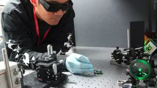
Researchers have developed a approach to exceed this limitation of the normal endoscopy methodology. They’ve created a photoacoustic imaging endoscopy probe that may match inside a needle with a diameter of 0.6mm. This gadget can resolve subcellular-scale tissue structural and molecular info in actual time 3D pictures. This information expertise permits a medical practitioner to determine and characterize the tissue in the course of the process.
Photoacoustic imaging makes use of shining pulses of sunshine with acoustic waves. The sunshine is concentrated on the constructions within the physique and the ultrasound sensors detect the acoustic waves to type 3D pictures for structural and useful info for scientific functions. Though this expertise requires a cumbersome ultrasound detector and has low imaging pace, this problem has been resolved. Researchers mixed wavefront-based beam shaping with light-based ultrasound detection and a quick algorithm for controlling the gadget. This distinctive mixture allowed them to create a particularly small probe with out sacrificing imaging pace.
The gadget consists of two optical fibers for delivering photoacoustic waves and one other for detecting ultrasound waves. A excessive pace digital micro-mirror with practically a million tiny mirrors is used for mild to scan exactly.
To exhibit the brand new gadget, the researchers used it to accumulate high-resolution pictures of mouse purple blood cells overlaying an space 100 microns in diameter. “We had been capable of accomplish this at about 3 frames per second,” Dr Xia mentioned. “We additionally confirmed that the needle probe might be scanned to considerably enlarge the field-of-view in real-time by stitching collectively consecutive pictures.”
The expertise has a broad vary of purposes, similar to fluorescent imaging, Raman microscopy and two-photon microscopy. “It may ultimately permit 3D characterization of tissue throughout varied minimally invasive procedures similar to tumor biopsies. This might assist clinicians pinpoint the best space to pattern, which might enhance the analysis accuracy.”
